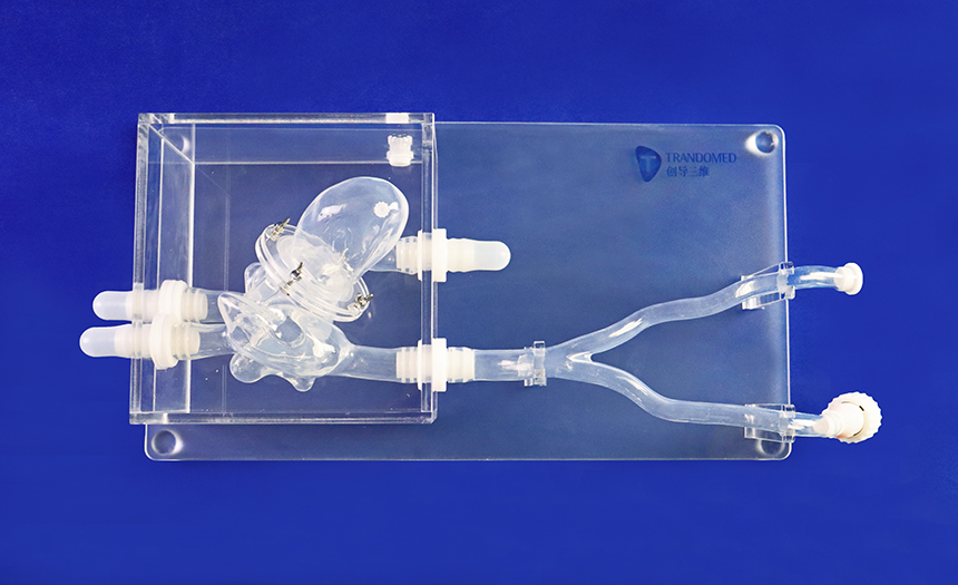
Mitral Regurgitation (MR), also known as mitral valve insufficiency or mitral valve insufficiency, is a valvular heart disease in which the mitral valve cannot close properly when the heart pumps blood. It refers to the abnormal phenomenon that blood leaks backward from the left ventricle through the mitral valve into the left atrium when the left ventricle contracts. MR is the most common form of valvular heart disease.
Symptoms and signs
Mitral regurgitation may be present for many years before any symptoms appear. The symptoms associated with MR depend on where in the disease process the patient is.
Patients with acute MR are often severely symptomatic, with signs and symptoms of acute decompensated congestive heart failure (i.e., tachypnea, pulmonary edema, orthopnea, and paroxysmal nocturnal dyspnea), as well as symptoms of cardiogenic shock (i.e., Shortness of breath at rest). Cardiovascular failure with shock (cardiogenic shock) may occur in individuals with acute MR, often due to papillary muscle rupture, villous tendon rupture, or mitral valve infective endocarditis.
Patients with chronic compensated MR may remain asymptomatic for long periods of time, have normal exercise tolerance, and have no evidence of heart failure. However, over time, it may decrease and the patient may develop volume overload (congestive heart failure). Symptoms of the decline may include fatigue, shortness of breath, especially with exertion, and swelling of the legs. In addition, an irregular heart rhythm called atrial fibrillation may occur .
The results of clinical examination depend on the severity and duration of MR. The mitral valve component of the first heart sound is usually soft, accompanied by a laterally displaced apex pulse, often with ups and downs. After the first heart sound, a high-pitched holosystolic murmur appears at the apex, radiating to the rear or clavicle area. Its duration, as the name suggests, is the entire systolic period. The loudness of the murmur has little to do with the severity of the reflux. This may be followed by a loud, palpable P2, which is best heard when lying on the left side, and the third heart sound is often heard.
In acute cases, murmurs and tachycardia may be the only obvious signs. Patients with mitral valve prolapse may have a holosystolic murmur or often a mid- to late-systolic and late-systolic murmur. Cases with late systolic regurgitant murmur may still be associated with significant hemodynamic consequences.
Pathophysiology
Mitral regurgitation is divided into acute (Acute MR) and chronic (Chronic MR) according to the development of the disease. Chronic regurgitation is also divided into organic (Degenerative MR, DMR/Primary MR) and functional (Functional MR, FMR/Secondary MR). ).
In clinical practice, the Carpentier classification, which is named based on the characteristics of valve disease, is often used: Type I has normal valve motion, that is, the valve itself has no disease or the disease is mild, mainly due to secondary annular dilation , such as secondary to dilated cardiomyopathy. Functional mitral regurgitation. Type II valve excessive movement is more common in patients with valve prolapse caused by long and ruptured chordae tendineae. Type III refers to limited valve movement, which is common in rheumatic heart valve damage, valve leaflet calcification caused by degeneration (type IIIa), and ischemic cardiomyopathy ( type IIIb ).
Acute mitral regurgitation is often caused by damage to different parts of the mitral valve structure. For example, infective endocarditis can cause valve leaflet perforation or chordae tendineae rupture, and patients with fibromatous mitral valve disease may develop spontaneous chordae tendineae rupture. Papillary muscle rupture may occur in patients with acute ST-segment elevation myocardial infarction, often accompanied by inferior wall infarction. Acute volume overload of the left atrium and left ventricle results in pulmonary congestion and low forward cardiac output. Timely diagnosis and emergency intervention of acute MR may be life-saving.
The two types of chronic mitral regurgitation actually have more differences in their presentation than they have in common. Organic lesions are often caused by diseases of the mitral valve device, while functional lesions are caused by lesions in the atria or ventricles. In chronic DMR, abnormalities in any valve component structure (valve leaflets, chordae tendineae, papillary muscles, valve annulus) can lead to valvular insufficiency and systolic blood pressure regurgitation from the left ventricle to the left atrium. In high-income countries, chronic DMR has a wide range of causes and manifestations, with mitral valve prolapse being the most common cause. Younger people often present with severe myxomatous degeneration, increased leaflet area, and lengthy chordae tendineae (Barlow 's valve). Some patients may develop ventricular arrhythmias, mitral annular disruption (figure), and left ventricular dilation. However, in older adults with fibroelastosis, rupture of the chordae tendineae may occur due to a lack of connective tissue. The distinction between these two etiologies may have implications for surgical intervention. Other less common causes of chronic DMR include infective endocarditis, connective tissue disease, rheumatic heart disease, mitral valve tears, and radiation heart disease. If volume overload is severe and sustained over a long period of time, it may cause myocardial damage, heart failure, and ultimately death. Reflux can be cured by correcting it before irreversible changes occur.
